-
CT scan protocol for patient-specific bone strength prediction
Download the CT Scan Protocol (PDF)
Calibration
At the outset of the study, European spine phantom (ESP) needs to be scanned on each CT scanner that will be used during the study. If such phantom is not available at the requester's hospital, one of our experts can do a calibration session on site with our own phantom. A calibration protocol should be performed for each scanner and for each Current/Voltage setting to be used in the study.
We assume that modern CT scanners x-ray tubes are stable for at least 12 months. If the study last longer than 12 months, then ESP scan should be repeated every 12 months.
Please note that this calibration is different from those performed by standard Quality Assurance protocols in place in most hospitals and is an essential requirement for model development. If such calibration cannot be carried out using a phantom, the user is encouraged to discuss with the team potential alternative options using phantomless approaches.
Imaging Protocol
All subjects should receive a bilateral full femoral CT scan, which should extend from 2 cm above the femoral head to 2 cm below the knee. In order to have an optimal calibration with an acceptable radiation dose the CT scans should be performed at 100 kV, and a constant tube current; any automatic algorithm that adjust current/voltage dynamically during the scan should be excluded.
We recommend using a standardised current of 120 mA, which is okay for female patients of average body weight. If the cohort includes patients with lower or higher than average body weight, we can process also data collected at 80, 100, 120, 140 mA, as far as the current remains constant during the whole scan. If this second approach is to be used, we will need a full calibration scan of ESP at each current value to be used in the cohort.
For GE CT scanners, we recommend using Standard reconstruction algorithm. For other CT scanners (Siemens, Toshiba etc) please contact us to discuss the appropriate reconstruction algorithm for the study.
Old CT Scanners
- Voltage
- 100 kV
- Current
- 120 mA
CT scan protocol should be organised in three blocks with different settings in the femoral diaphysis and the two epiphyses. For spiral CT systems the Pitch P and the longitudinal dimension of the detector collimation D should be set for each block as shown below.
Old CT scanner settings: voltage 100 kV, current 120 mA Region Pitch (P) Detector collimation (D) Slice thickness (S) Proximal epiphysis 2 3 2 mm Diaphysis 2 5 4 mm Distal epiphysis 2 3 2 mm New CT Scanners
- Voltage
- 100 kV
- Current
- 120 mA
CT scan should be performed with uniform slice thickness of 0.625 mm and a pitch of 1 over the whole femur.
The scan-region breakdown used on older scanners (2 mm slice thickness in the proximal and distal epiphyses, 4 mm in the diaphysis) is illustrated in Figure 1 of the downloadable PDF above.
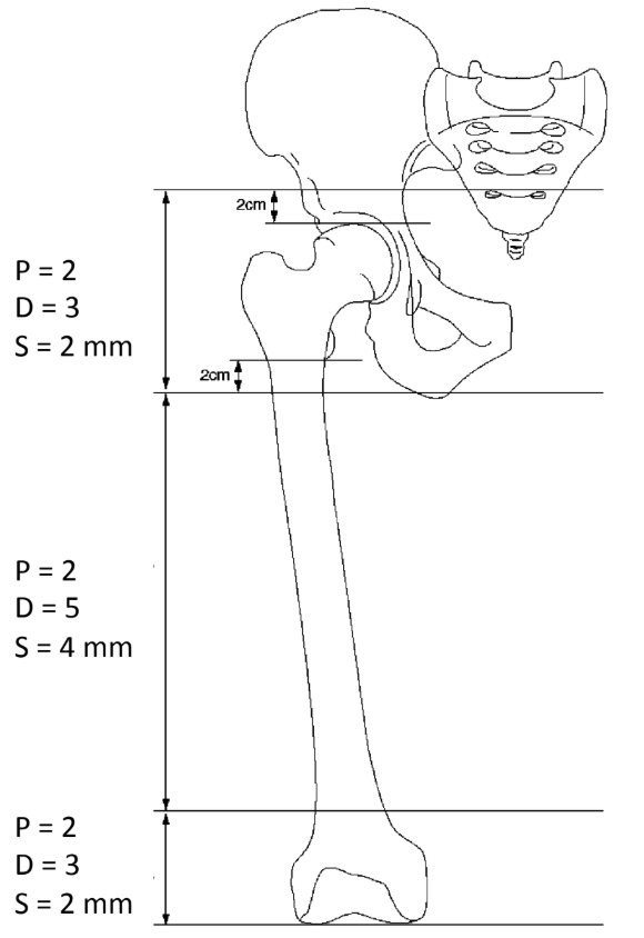
Whole femur should be scanned with a slice thickness of 2mm in the distal and proximal epiphysis and 4 mm in the diaphysis. Radiation Dose
The ImPACT CT Patient Dosimetry Calculator was used to compute effective radiation dose. It makes use of the “Normalised organ doses for x-ray computed tomography calculated using Monte Carlo techniques” NRPB-SR250 released by Public Health England on 11/2014, and based on the organ weighting scheme ICRP-103. It calculates the effective dose in mSv for each organ present within the scan length. Mathematical phantom used with ImPACT software extends from head through proximal femur only. CT dose calculations were done for GE LightSpeed CT scanner. Effective dose was calculated for a proximal femur scan in males and females.
Currently the proximal femur scan is performed at 120 kV and the tube current is modulated between 80 mA and 200 mA with a slice thickness of 0.625mm. This corresponds to effective dose in the range of 2.5–6.2 mSv for males and 1.5–3.80 mSv for females.
When we analyse the distribution of dose in different organs, we find that majority of the dose is contributed by gonads, colon and bladder for both females and males. For example, the total effective dose for males at 120 kV/200 mA is 6.2 mSv. Its distribution among different organs in mSv is as follows:
Effective dose by organ, males at 120 kV/200 mA (mSv) Organ Dose (mSv) Gonads 3 Colon 1 Bladder 1.2 Bone marrow 0.47 Bone surface 0.09 Skin 0.06 - Voltage
- 120 kV
- Current
- 80–200 mA (modulated)
- Slice thickness
- 0.625 mm
Thus, more than 80% of the dose is contributed by the above-mentioned three organs. If we extend the scan length to include whole femur up to below the knee, the additional dose contribution would come from bone marrow, bone surface and skin. In order to estimate the effective dose for full femur scan, we assumed the additional dose to be double the value of total dose contributed by bone marrow, bone surface and skin in the proximal femur scan. For the current clinical protocol settings, it will be an additional 1.24 mSv.
By reducing the tube voltage from 120 kV to 100 kV while keeping the remaining scan settings the same, the whole femur scan would result in an effective radiation dose in the range of 1.9–4.8 mSv for males and 1.3–3.2 mSv for females.